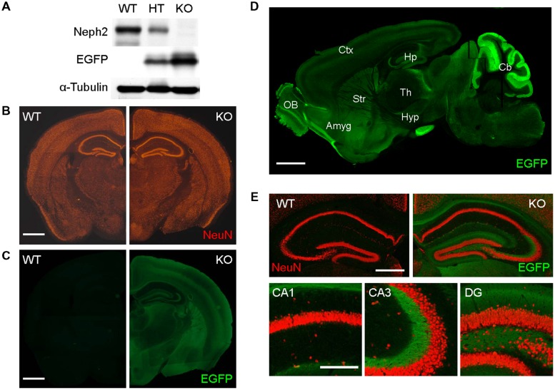
FIGURE 1.
Normal gross morphology of the Neph2-/- brain. (A) Absence of Neph2 protein detection in the Neph2-/- brain. WT, Neph2+/- (HT), and Neph2-/- (KO) whole brain lysates (3 weeks) were immunoblotted with Neph2 (1468 rabbit) antibodies. Note that EGFP expression driven by the endogenous Neph2 promoter is higher in the Neph2-/- brain, relative to Neph2+/- brain. (B) Normal gross morphology of the Neph2-/- brain (3 weeks), revealed by NeuN (neuron-specific marker) staining. Scale bar, 0.5 mm. (C–E) Expression patterns of Neph2 in the brain, revealed by EGFP expression driven by the endogenous Neph2 promoter. Neph2-/- coronal (C,E) and sagittal (D) brain sections (3 weeks) were doubly stained for EGFP and NeuN. (E) Shows further details of Neph2 expression in the hippocampal coronal section. Ctx, cortex; Hp, hippocampus; Str, striatum; Th, thalamus; OB, olfactory bulb; Amyg, amygdala; Hyp, hypothalamus; Cb, cerebellum. Scale bar, 0.5 mm for (B–D) and 0.2 mm for (E).

