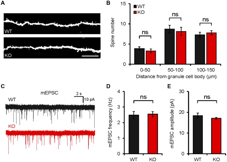
FIGURE 6.
Neph2-/- mice show normal spine density and spontaneous excitatory transmission in the hippocampal DG. (A,B) Normal density of dendritic spines in Neph2-/- DG granule cell dendrites (∼8 weeks), as measured by biocytin infusion into Neph2-/- slices. (n = 11 cells from 5 mice for WT, and 14 cells from 5 mice for KO, ns, not significant, Student’s t-test). (C–E) Normal frequency and amplitude of mEPSCs in Neph2-/- DG granule cells (8 weeks). (n = 16, 5 for WT, and 17, 5 for KO, ns, not significant, Student’s t-test).

