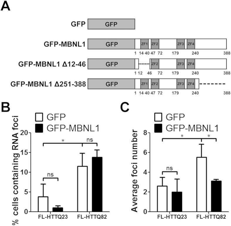
Figure 3. MBNL1 decreases the number of FL-HTTQ82 RNA foci.
(A) Schematic representation of GFP-MBNL1 plasmids. ZFs, zinc finger motifs. SK-N-MC cells were co-transfected with FL-HTT and GFP-MBNL1 plasmids for 48 hours and subjected to FISH. GFP plasmid was used as a control. Foci analysis was performed by Nikon Eclipse E400 microscopy. In each treatment, numbers of GFP-positive cells and foci-containing GFP-positive cells were counted. (B) Percentage of cells containing RNA foci. (C) Average foci number. Both experiments, two-way ANOVA, n = 3 biological replicates; *P < 0.05, **P < 0.01, ns = no significance.

