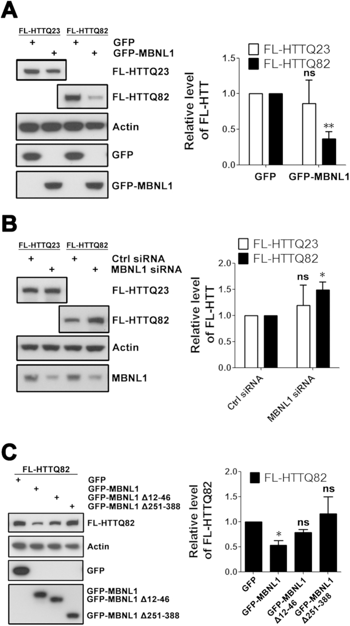
Figure 5. MBNL1 decreases expression of expanded FL-HTT protein.
(A) SK-N-MC cells were co-transfected with FL-HTT and GFP-MBNL1 plasmids, and levels of FL-HTT protein were assessed by western blot 72 hours post-transfection. GFP plasmid was used as a control. Overexpression of MBNL1 decreased levels of FL-HTTQ82. Student’s t-test, n = 3 biological replicates. **P < 0.01, ns = no significance, versus GFP group. (B) SK-N-MC cells were first transfected with MBNL1 siRNA and FL-HTT plasmid. Levels of FL-HTT were assessed by western blot. Control siRNA was used as a control. Knock-down of endogenous MBNL1 increased expression of FL-HTTQ82. Student’s t-test, n = 3 biological replicates. *P < 0.05, ns = no significance, versus control siRNA group. (C) SK-N-MC cells were co-transfected with FL-HTT and GFP-MBNL1 plasmids, and levels of FL-HTT protein were assessed by western blot 72 hours post-transfection. GFP plasmid was used as control. Overexpression of GFP-MBNL1 Δ12–46 (loss of first zinc finger) and GFP-MBNL1 Δ251–388 (loss of C-terminal splicing domain) abolished the effect of MBNL1 on FL-HTTQ82 levels. One-way ANOVA, n = 3 biological replicates. *P < 0.05, ns = no significance, versus GFP group.

