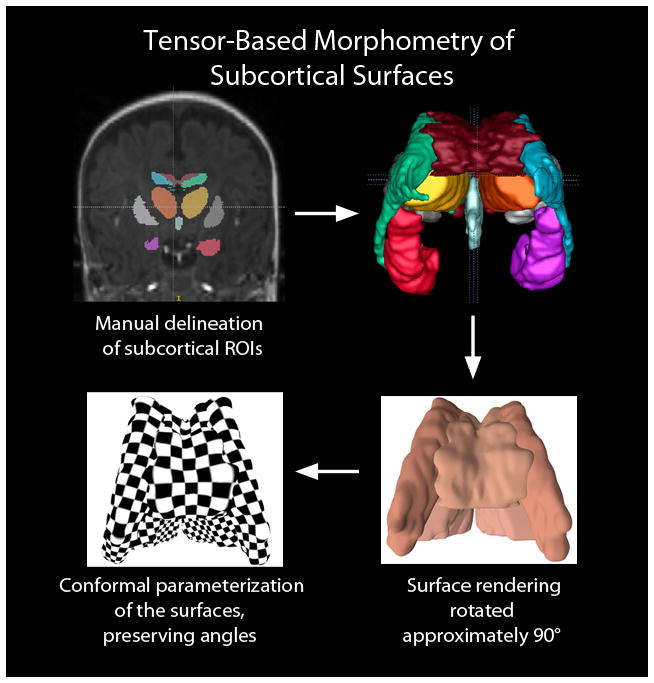
Fig. 11.

Application of tensor-based surface morphometry to the subcortical structures of a developing preterm infant brain derived from coronal SPGR 3D-volumedatasets. The first step in data processing (top left) involves manual delineation of subcortical structures. From there, 3D surfaces can be generated (top right) representing the corpus callosum (dark red), lateral ventricles (green and blue, representing the left and right lateral ventricles, respectively), hippocampus (orange red and purple, representing the left and right hippocampi, respectively), and third ventricle (light blue). The putamen, which were manually delineated in the coronal SPGR have not been included in this 3D rendering. The bottom left illustrates the computed conformal parameterization of the surfaces. The checkerboard texture map shows the angle-preserving property (right angles on the plane are mapped to right angles on surfaces). The surface conformal parameterization provides an ideal canonical space to register subcortical structures and analyze their interaction-related morphometry changes
