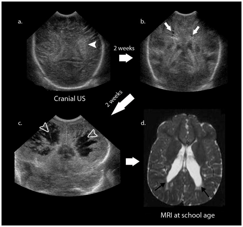
Fig. 2.

The classic evolution of cystic periventricular leukomalacia on cranial US. The first image (a) shows diffuse increased echogenicity (white arrowhead) followed serially by two other studies 2 weeks apart (b, c), which show the development of hypoechoic cystic leukomalacia (white arrows) and severe cavitary periventricular leukomalacia (open white arrowheads). The longterm sequelae of these neonatal findings are cerebral palsy and spastic diplegia. d Axial T2-W MR image during follow-up examination of this patient at school-age shows enlargement of the lateral ventricles at the level of the trigone extending caudally (black arrows) with adjacent region of periventricular T2 hyperintensity
