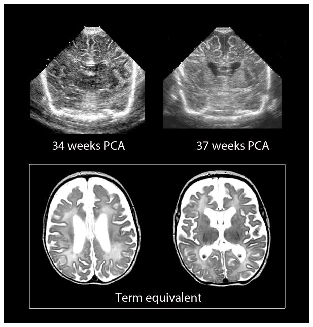
Fig. 4.
Chronic diffuse white matter injury in the preterm infant can be detected by both cranial US and MRI. Images of serial cranial US performed at 34 and 37 weeks’ gestation (top) show progressive enlargement of the lateral ventricles and concomitant increases in the sulci and subarachnoid spaces. The corresponding MR performed at term-equivalent age (bottom) shows similar findings likely related to diffuse white matter hypoplasia

