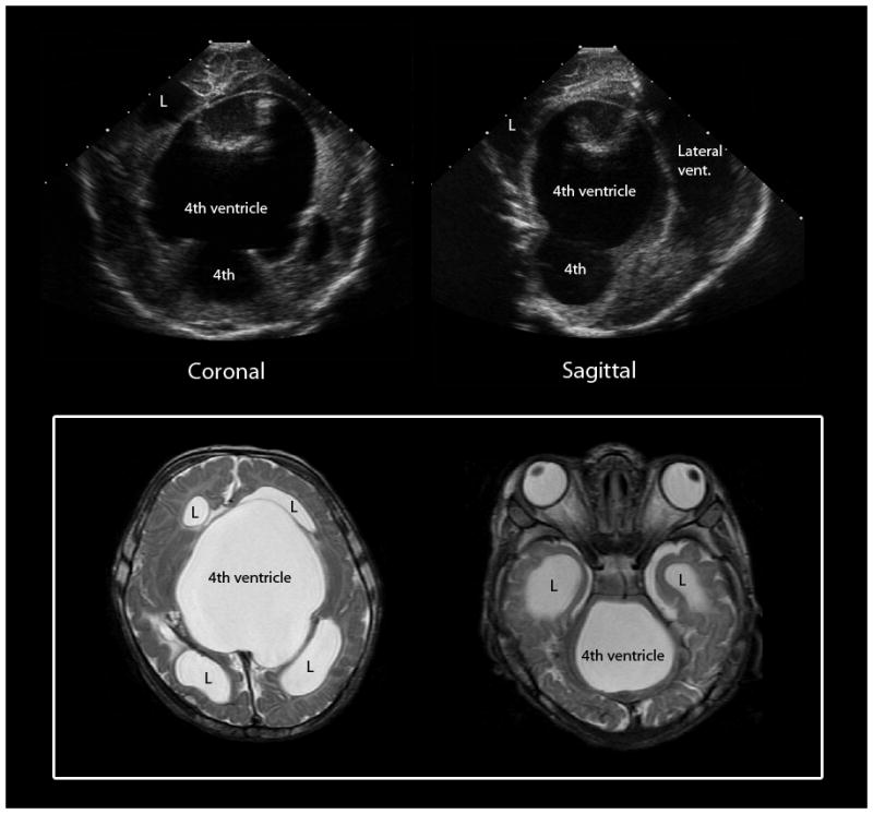
Fig. 5.

Trapped 4th ventricle, a complication of post-hemorrhagic hydrocephalus. Cranial US images (top) show a large cystic structure extending from the posterior fossa supratentorially through the tentorial incisura. Axial T2-weighted MR images (bottom) confirm an enlarged 4th ventricle with blocked outlet at the level of the aqueduct of Sylvius and the bilateral foramen of Lushka. L = lateral ventricle
