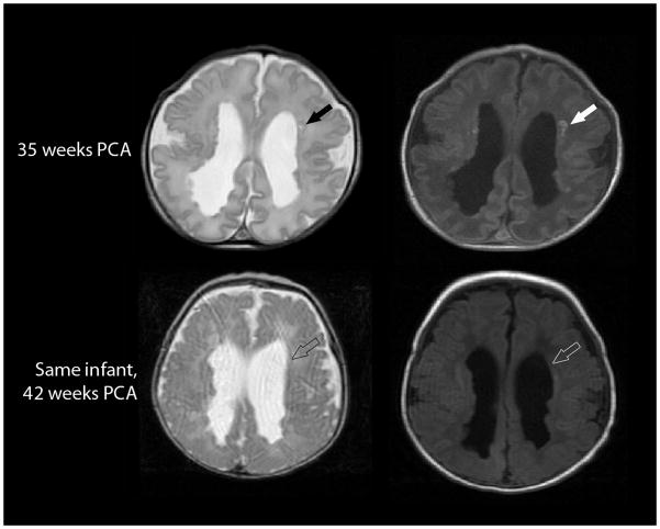
Fig. 7.
Evolution of a focal non-cystic PVL on axial T2-weighted (left) and axial T1-weighted (right) MRI in a 34-week gestation preterm infant imaged at 35 weeks postconceptional age (top row) and 42 weeks postconceptional age (bottom row). The T1 hyperintense punctate non-cystic lesions evidenced on the early scan (solid arrows) could not be visualized on the later scan (open arrows). (Of note, these studies also demonstrate numerous nodules along the surface of the lateral ventricles, presumed to be heterotopias.)

