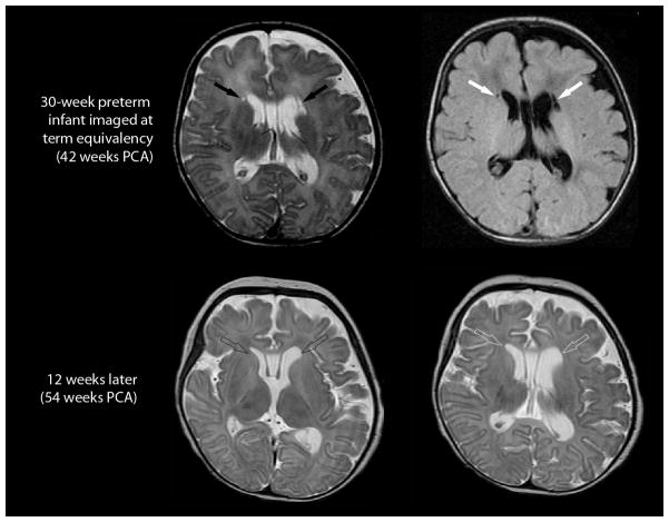
Fig. 8.
Evolution of a focal cystic PVL in an infant born at 30 weeks PCA. Imaging at term-equivalency (42 weeks PCA; top row, axial T2-weighted image on the left and axial FLAIR image on the right) demonstrates . . focal cystic periventricular lesions (solid arrows) abutting the frontal horns of the lateral ventricles. Comparable cuts on a follow-up MRI conducted 12 weeks later (bottom, both T2-weighted sequences), shows no evidence of cystic lesions adjacent to the lateral ventricles (open arrows). Instead, the follow-up MRI shows enlargement of the lateral ventricles and associated white matter volume loss.

