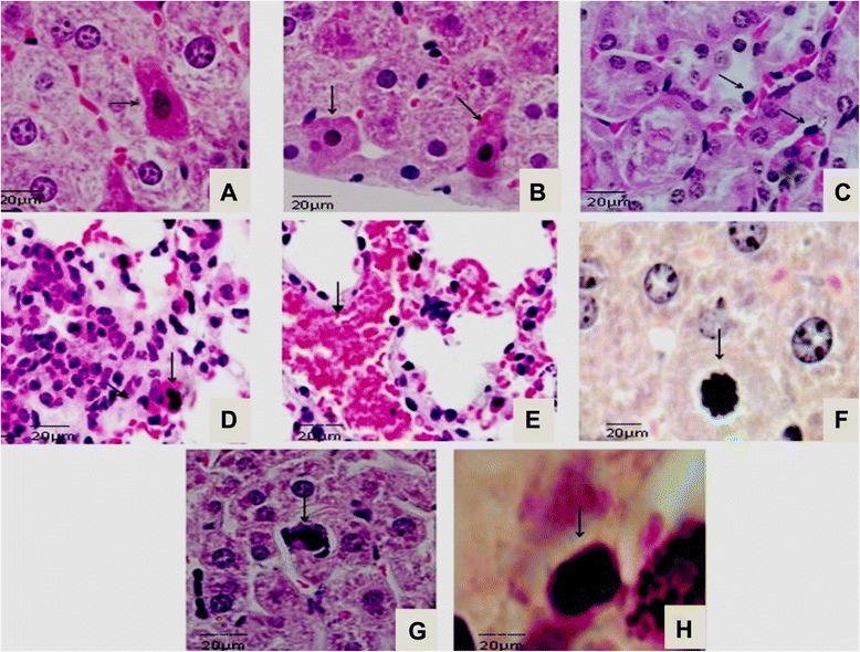
Fig. 1.

Histological lesions in BALB/c infected with L. interrogans serovar Icterohaemorrhage. Shown are sections of (a and b) liver, (c) kidneys, and (d and e) lungs. Hepatocytes showing eosinophilic cytoplasm and nuclear condensation at 14 dpi (arrows); kidneys degeneration followed by acute tubular necrosis in cortical tubules at 21 dpi (arrow); lung tissue at 14 dpi showing diffuse inflammatory cell infiltrate close to hilum region (arrow); condensation of chromatin and apoptotic bodies observed in kidney and liver sections (arrows) (n = 5 animals). Bar: 20 μm
