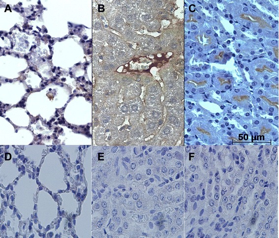
Fig. 2.

Immunodetection of caspase-3 reactive cells in BALB/c lung, liver and kidney tissue infected with L. interrogans serovar Icterohaemorrhage. a Lung samples at 14 dpi revealed positive signals in the alveolar epithelium. b Liver samples at 14 dpi showed centrilobular vein endothelium reactive to caspase-3 antigen. c In the kidney, at 21 dpi, tubular epithelial cells positively labeled for caspase-3 were observed. (d, e and f). Lung, liver and kidney respective controls (n = 5 animals)
