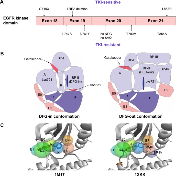
Figure 2.
Mutations in EGFR kinase domain and topological distribution of the binding pockets in the catalytic cleft.
Notes: (A) TKI-sensitive and resistant mutations. (B) Subregions of binding pockets of EGFR with DFG-in conformation and DFG-out conformation. A, adenine binding pocket; R, ribose pocket; P, phosphate pocket; E0, entrance pocket 0; E1, entrance pocket 1; E2, entrance pocket 2; K, small region in the deep front pocket; BP-I, back pocket I; BP-II, back pocket II; BP-III, back pocket III; BP-IV, back pocket IV. (C) Mapping the ligands (Erlotinib in 1M17 EGFR protein and lapatinib in 1XKK EGFR protein) into subregions of the binding pockets in the 3D view.
Abbreviations: EGFR, epidermal growth factor receptor; TKI, tyrosine kinase inhibitor; BP, back pocket; LREA, Leu-Arg-Glu-Ala; NPG, Asn-Pro-Gly; SVQ, Ser-Val-Gln; DFG, Asp-Phe-Gly.

