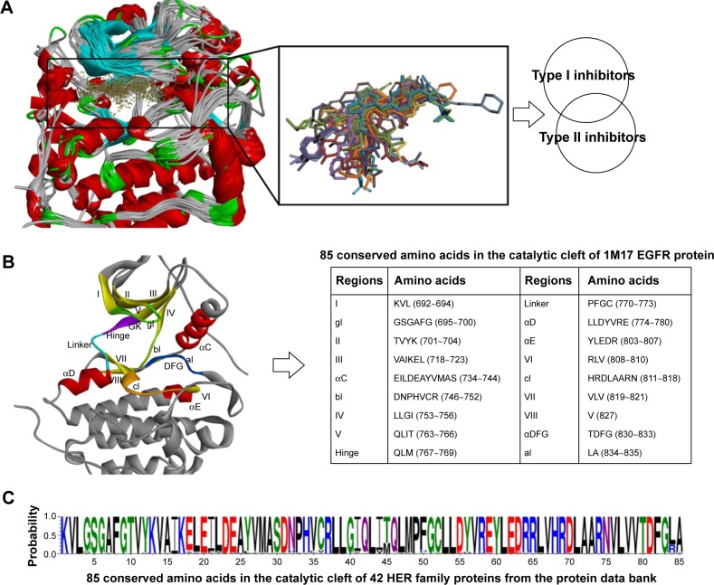
Figure 3.
Analysis of binding pockets extracted from 42 HER family proteins with small-molecule ligands.
Notes: (A) Alignment of these 42 tyrosine kinase domains using the Discovery studio 3.5 based on the sequence similarity. (B) Structure of the EGFR kinase domain, with important structural elements labeled according to KLIFS database (I–VIII = β-sheets I–VIII; linker = loop connecting the hinge to αD-helix). (C) Conservation of the 85 amino acids in the catalytic cleft of 42 HER kinase inhibitor complexes. This picture was drawn by WebLog 3.4.
Abbreviations: gl, G-rich loop; bl, loop connecting αC-helix to IV; GK, gatekeeper; cl, catalytic loop; DFG, Asp-Phe-Gly; xDFG, DFG-motif plus one preceding amino acid residue; al, activation loop.

