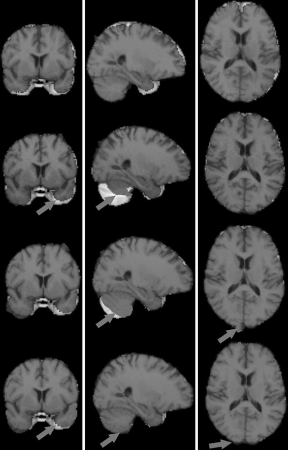
Fig. 2.

Visual results for the reference image, input image and two normalized images in the same slice for one subject registered with the brain template image MNI152-2mm_brain. From top to bottom: reference image, input image, and images normalized with Hist. Matching and Hist. Normalization overlaid with template image. From left to right: coronal slice, sagittal slice, axial slice. The arrow points to the differences of visual results of registration between images and brain template image.
