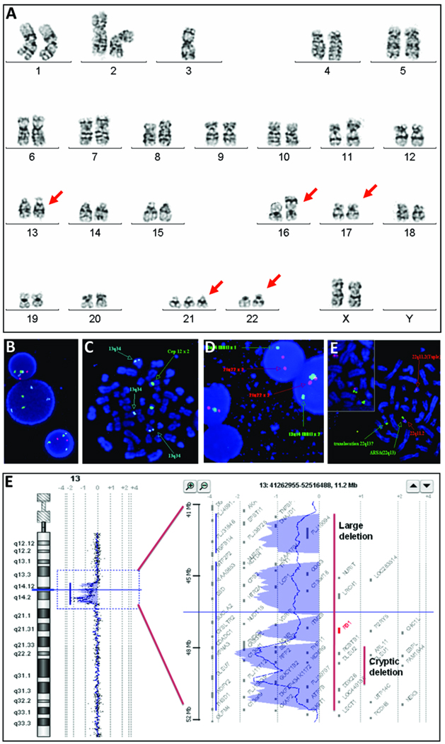
Figure 1.
Karyotype and FISH analysis on cultured unstimulated bone marrow cells. A: Chromosomal spread reveals a clonal abnormality detected at initial diagnosis including a 13q deletion, der (16), t(17;22) and acquired trisomy 21. B: Fish analysis on interphase cells reveals loss of 13q14.3 (red). C: FISH analysis on metaphase spread demonstrates homozygous loss of 13q14 (red) and the presence of 13q34 (aqua) on chromosome 16. D: Fish analysis on interphase cells using probes for RB1 gene and 21q22.13 (red) reveals only one copy of the RB1 gene in two cells. E: Fish analysis on metaphase spread identifies a translocation between 17q and 22q with the presence of the 22q13 signal (green) in the der(17) and the 22q11.2 signal (red) in the der(22). F: oaCGH analysis performed at time of first relapse defines a 1.72 Mb deletion of 13q14.2-q14.3 and a 9.34 Mb deletion of 13q14.11-q14.3.

