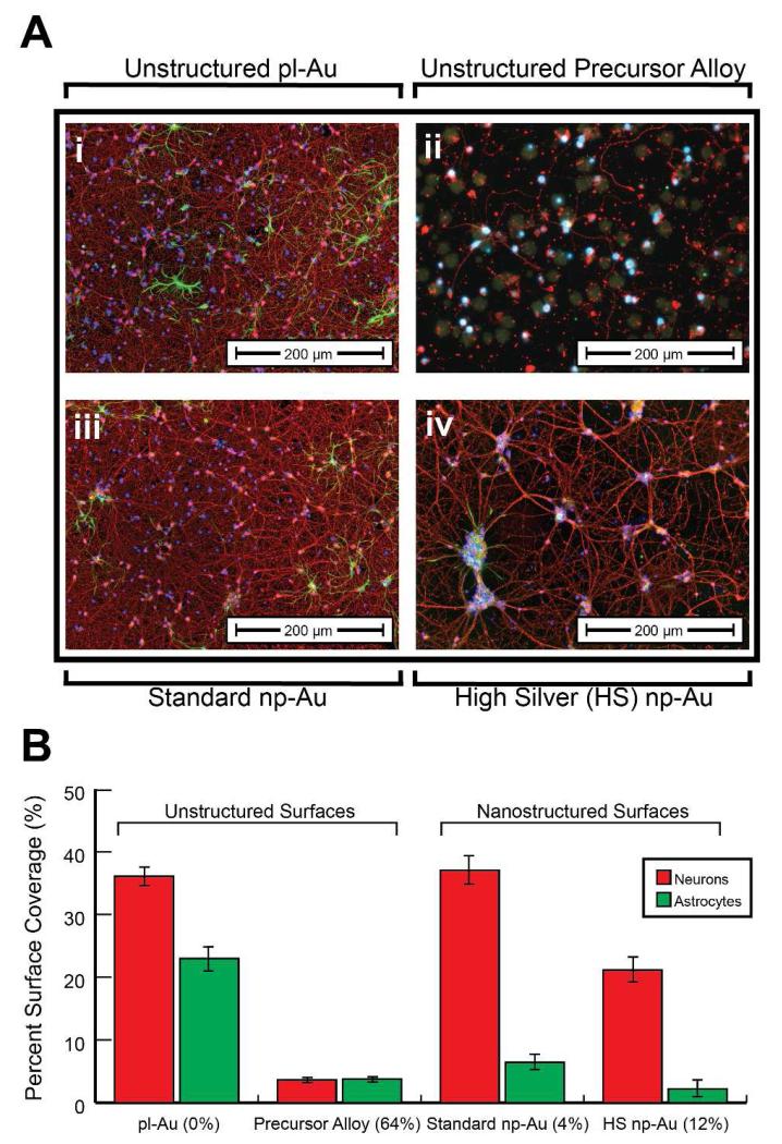
Figure 3.
(A) Merged epi-fluorescent images of primary cortical neuron-glia co-cultures immunostained for β-tubulin (red – neurons) and GFAP (green – astrocytes) grown on unstructured pl-Au (i) and unstructured precursor gold-silver alloy with 64% silver (ii), as well as standard np-Au containing ~4% silver (iii) and high silver content np-Au film containing ~12% silver (iv). (B) Surface coverage analysis of day in vitro 7 neurons and astrocytes grown on the np-Au films containing varying amounts of silver (12% and 4%), as well as pl-Au and the gold-silver alloy reveals acute toxicity of the high silver np-Au with no visually observed toxicity of the standard np-Au.

