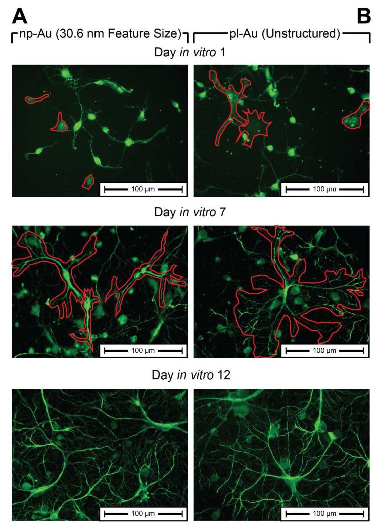
Figure 9.
High magnification (40×) images of neurons and astrocytes at DIV 1, DIV 7, and DIV 12 on (A) standard np-Au and (B) unstructured pl-Au reveal cellular differences between astrocyte growth on np-Au and pl-Au. Astrocytes are highlighted by a red outline in DIV 1 and DIV 7 images. Due high non-specificity of glial fibrillary acidic protein in perinatal neurons and astrocytes, neurons are visible in DIV1 stains.51 Astrocytes and neurons were differentiated visually through a co-localization of tubulin-β-III and GFAP (not shown here).

