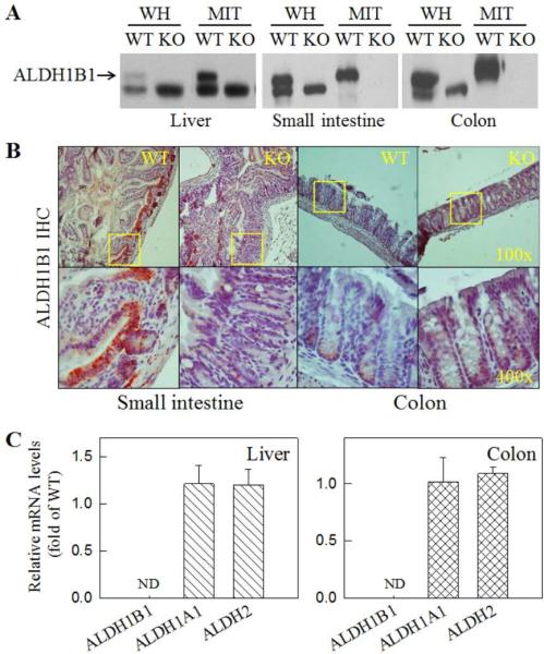
Fig. 1. Loss of ALDH1B1 expression in ALDH1B1 KO mice.
(A) Western blotting detection of ALDH1B1 protein in whole-cell homogenates (WH) and mitochondrial extractions (MIT) from liver (10 g), small intestine (10 g) and colon tissues (40 g). The rabbit polyclonal anti-ALDH1B1 antibody appeared to cross-react with an unknown lower molecular weight peptide. (B) Immunohistochemical (IHC) detection of ALDH1B1 in mouse small intestine and colon tissue sections. Cells at the crypt base showed ALDH1B1 immunoreactivity in WT but not in KO mice. Representative images are presented at two magnifications, with the square field in the top panel (100x) enlarged in the bottom panel (400x). (C) qRT-PCR detection of mRNAs of ALDH1B1, ALDH1A1 and ALDH2 in liver and colon tissues. Relative mRNA levels were expressed as fold of control (WT = 1) after normalization with β-2 microglobulin (B2M). Data are presented as mean + SE from 4 mice. ND, not detectable.

