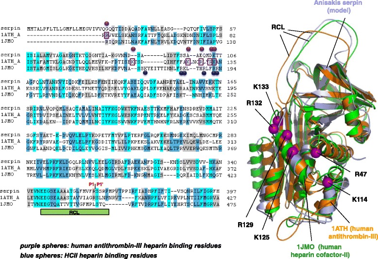
Fig. 5.

Sequence and structural alignment of ANISERP, human antithrombin III (1 ATH_A), and human heparin cofactor-II (1JMO). The key residues involved in heparin binding described for human antithrombin III (Arg 47, Lys114, Lys125, Arg 129, Arg132 and Lys133) are shown as purple boxes and spheres. The key residues involved in heparin binding described for heparin cofactor-II (Lys173, Arg184, Lys185, Arg189, Arg192 and Arg193) are shown as blue spheres. The positions of the P1-P1’residues in the ANISERP sequence are indicated. The residues in the 1JMO and 1ATH_A sequences are numbered according to Baglin et al. [26]
