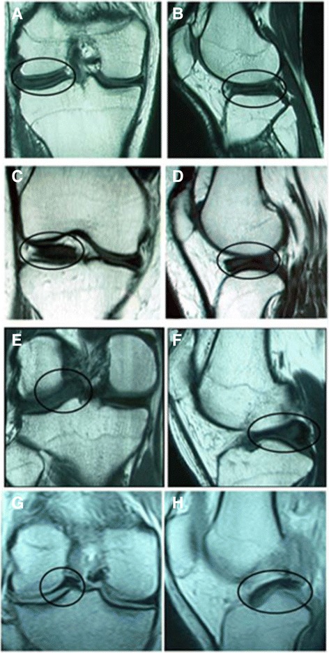
Fig. 1.

T1-weighted MR images of DLM with different types of shifts. Coronal (a) and sagittal (b) images of no shift, coronal (c) and sagittal (d) images of anterocentral shift, coronal (e) and sagittal (f) images of posterocentral shift and coronal (g) and sagittal (h) images of central shift
