Fig. 1.

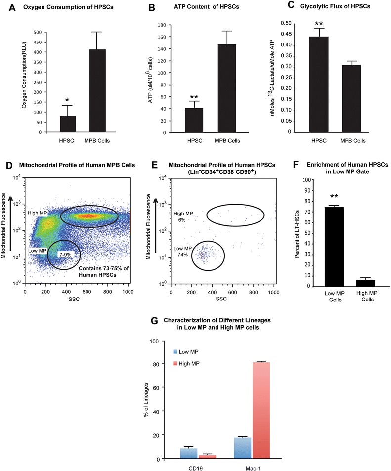
Metabolic profile of human HPSCs. a) Oxygen consumption of Lin−CD34+CD38−CD90+ HPSCs and human mobilized peripheral blood cells (MPB Cells) demonstrating lower rates of oxygen consumption by HPSCs (n = 3). b) ATP level of HPSCs and MPB Cells demonstrating lower ATP levels in HPSCs (n = 3). c) Glycolytic flux of HPSCs and MPB Cells demonstrating higher rates of glycolysis in HPSCs (n = 3). d) Flow cytometry profile of MPB mononuclear cells stained with mitotracker. Note populations with different mitotracker fluorescence. e) Mitotracker profile of human HPSCs. The majority of human HPSCs (73–75 %) are localized to a unique population (7–9 %) of total human MPB mononuclear cells with low mitochondrial potential (low MP). f) Quantification of the percent of human HPSCs residing in low MP cells and high MP cells demonstrates significant enrichment of human HPSCs in low MP gate. g) Characterization of different lineages within low and high MP cells of MPB. The low MP cells only contained a few B cells (CD19, 7.9 %) and myeloid cells (Mac-1, 17.05 %). The high MP cells had lower percentage of B cells (CD19, 2.4 %), but much higher percentage of myeloid cells (Mac-1, 81.65 %) (n =3).*p < 0.05, **p < 0.01
