Figure 3.

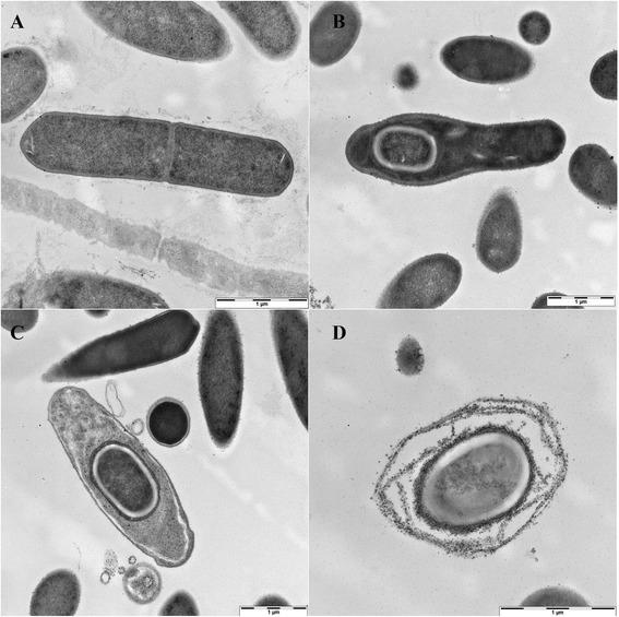
Transmission electron microscopy image of C. sporogenes DSM 795; A: dividing cell; B, C: sporulating cells; D: spore; scale bars represent 1 μm.

Transmission electron microscopy image of C. sporogenes DSM 795; A: dividing cell; B, C: sporulating cells; D: spore; scale bars represent 1 μm.