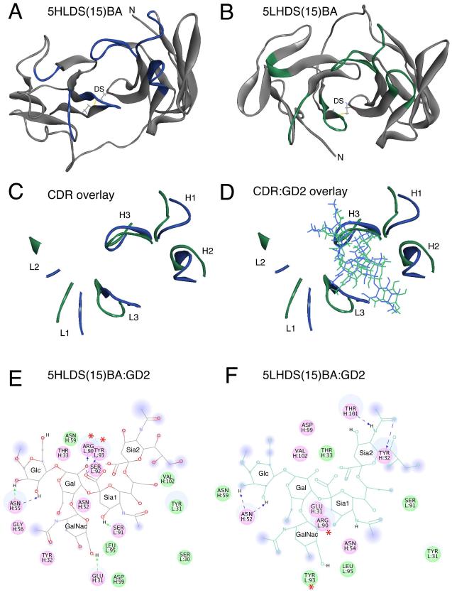
Figure 1. Molecular Modeling of anti-GD2 5F11-scFv in the 5HLDS(15)BA and 5LHDS(15)BA constructs.
Molecular Modeling of anti-GD2 5F11-scFv in the 5HLDS(15)BA and 5LHDS(15)BA constructs. A) Ribbon diagram of 5F11-scFv in 5HLDS(15)BA (5F11 VH - linker - VL - disulfide stabilized), where VH-VL disulfide stabilizing residues are shown in stick rendering, and CDR main chain residues are shown in Blue. B) Ribbon diagram of 5F11-scFv in 5LHDS(15)BA (5F11 VL - linker - VH - disulfide stabilized), where VH-VL disulfide stabilizing residues are shown in stick rendering, and CDR main chain residues are shown in Green. C) Overlay of CDR loops of 5HLDS (Blue) and 5LHDS (Green). D) Overlay of CDR residues with docked model of GD2 for 5HLDS(15)BA:GD2 (Blue) and 5LHDS15BA:GD2 (Green). E) Interaction diagram of 5HLDS(15)BA:GD2 and F) Interaction diagram of 5LHDS(15)BA:GD2. Interacting residues are displayed as colored discs. Residues with H-bonding, charged or polar interactions are colored in magenta. Residues having van der Waals interactions are colored in green. H-bonds, and charge-charge interactions are displayed as dashed lines. The solvent accessible surface is shown as a diffuse background circle with the radius proportional to the exposure. The per residue interaction energies are shown in Supplemental Table III. The residues with the largest contributions to the interaction with GD2 are L: Arg90 and L: Tyr93, which are significantly reduced in 5LHDS(15)BA:GD2 (noted with *).

