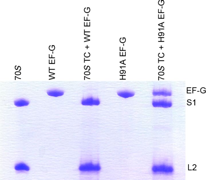
Fig. 2. Occupancy of H91A EF-G in the ribosomal translocation complex.
Translocation complexes (70S TC) containing WT or H91A EF-G are ultracentrifuged at 100,000g at 4°C with 30% sucrose cushion. The ribosomal pellet was carefully isolated, washed, and subjected to SDS-PAGE analysis. Whereas no EF-G band was seen in the 70S TC + WT EF-G complex, a clear band of H91A EF-G, in almost 1:1 stoichiometry with the ribosomal protein S1, could be seen in the 70S TC + H91A EF-G complex.

