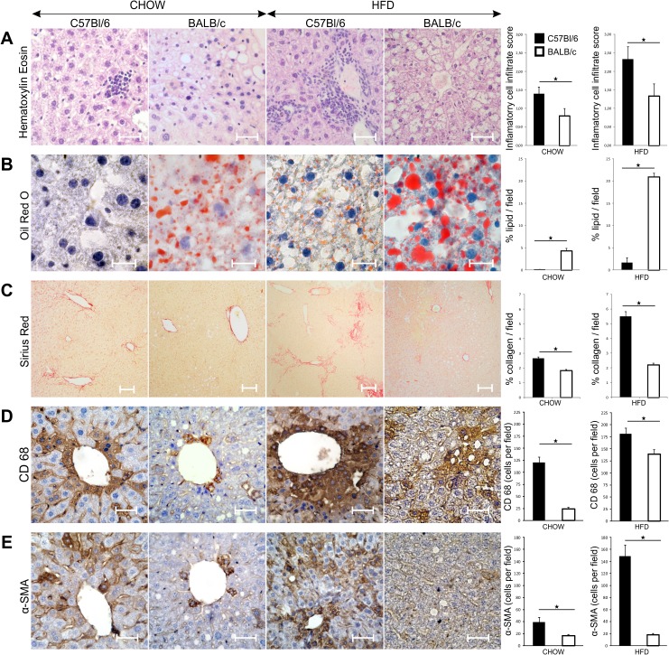
Fig 6. Liver inflammation, steatosis and fibrosis in chow and HFD-fed C57Bl/6 and BALB/c mice.
(A) Representative images of HE staining of paraffin-embedded liver tissue sections (original magnification x40, scale bar = 50μm). Results are presented as inflammatory infiltrate score according to NASH score system. Inflammatory infiltrate score was significantly higher in chow- and HFD-fed C57Bl/6 mice compared with diet-matched BALB/c mice at 32 weeks of age. (B) Representative images of Oil Red O staining on frozen liver sections (original magnification x100, scale bar = 25μm). Results are presented as percentage of red stained area relative to the total section area. Percentage of stained lipids was significantly lower in chow- and HFD-fed C57Bl/6 mice compared with diet-matched BALB/c mice at 32 weeks of age. (C) Representative images of Sirius Red staining on paraffin-embedded liver tissue sections (original magnification x10, scale bar = 100μm). Results are presented as percentage of red stained area relative to the total section area. Percentage of stained collagen fibers was significantly higher in chow- and HFD-fed C57Bl/6 mice compared with diet-matched BALB/c mice at 32 weeks of age. (D) Representative images of CD68 IHC staining on paraffin-embedded liver sections (original magnification x40, scale bar = 50μm). Number of CD68 positive cells per field was significantly higher in chow- and HFD-fed C57Bl/6 mice compared with diet-matched BALB/c mice at 32 weeks of age. (E) Representative images of αSMA IHC staining on paraffin-embedded liver sections (original magnification x40, scale bar = 50μm). Number of α-SMA positive cells per field was significantly higher in chow- and HFD-fed C57Bl/6 mice compared with diet-matched BALB/c mice at 32 weeks of age. The results are shown as the means ± SEM of 9–10 animals per group. *p<0.05. The results are representative of two experiments.

