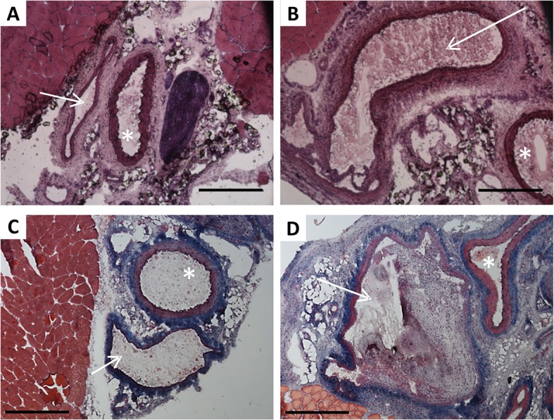
Fig 2. Femoral blood vessels morphology () in sham-operated limbs (A, C) and limbs with AVF (B, D).

Panels A, B—hematoxylin and eosin stained cryosections; panels C, D—Masson’s trichrome stained cryosections. White arrow indicates the vein, asterisk–the artery. Scale bar: 500μm
