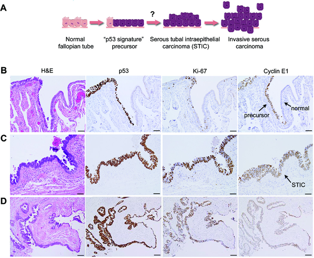
Figure 3. Cyclin E1 expression occurs early during serous tumorigenesis.
A, An illustration of the proposed carcinogenic sequence for serous tumorigenesis in the fallopian tube epithelium. The “p53 signature” is hypothesized to precede STIC development. B, Example of high Cyclin E1 expression in a putative precursor lesion. Precursor cells are differentiated from adjacent normal cells by their nuclear p53 expression (brown), typically indicating somatic mutation of TP53. Note that Cyclin E1 is expressed in precursor cells but not in adjacent normal cells. The proliferation marker Ki-67 is absent in most precursor cells, indicating low proliferative activity. H&E (hematoxylin and eosin) staining highlights histologic features. C-D, Example of high Cyclin E1 expression in STIC, shown at high (C) and low (D) magnification. STICs are highly proliferative, indicated by high Ki-67 expression, and typically express mutant p53. STICs usually exhibit nuclear atypia and loss of polarity, evident by H&E staining. Scale bars = 20 µm (B-C), 50 µm (E).

