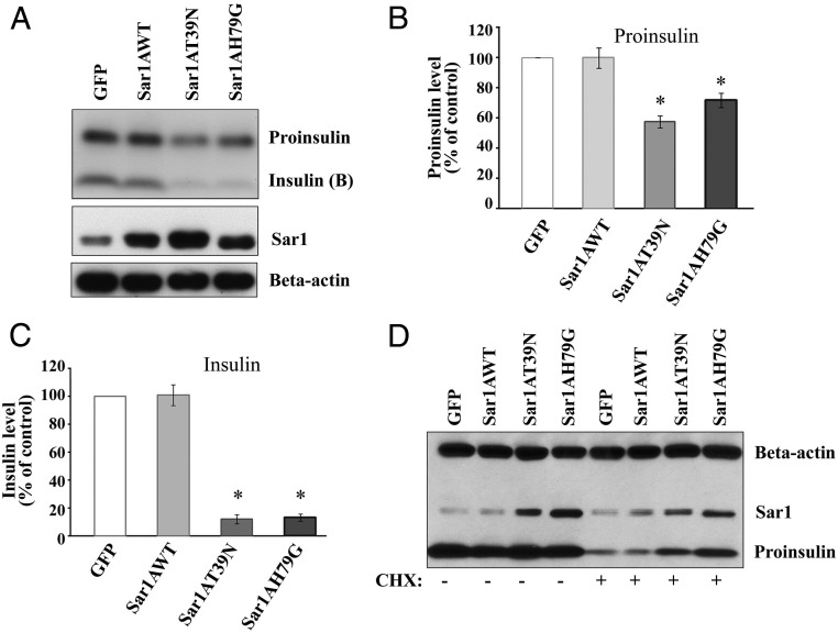
Figure 3.
Sar1 mutants blocked ER exit of endogenous proinsulin. A, MIN6 cells were infected with either GFP, wild-type Sar1A, Sar1AT39N, or Sar1AH79G adenovirus. After 48 hours, total protein lysates were harvested and analyzed by Western blot analyses with anti-insulin, anti-Sar1A, and antiactin antibodies. After normalized with β-actin, the protein levels of proinsulin (B) as well as insulin (B-chain) (C) were expressed as the percentage (mean ± SD, n = 4) of that of the control GFP virus infected MIN6 cells. *, P < .05. D, MIN6 cells were infected with either GFP, wild-type Sar1A, Sar1AT39N, or Sar1AH79G adenovirus. After 22 hours, infected cells were treated with or without CHX at 15 μg/mL for 2 hours. Then, the total protein lysates were harvested and analyzed by Western blotting with anti-insulin, anti-Sar1, and antiactin antibodies. A representative image of 3 separate experiments is shown.

