Figure 4.
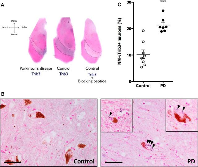
The proportion of Trib3+ dopaminergic neurons is increased in the substantia nigra of PD patients. A, Representative low-magnification images (4×) of postmortem midbrains from age-matched human control and PD patients immunostained for Trib3 (blue) and counterstained with Fast Red (pink) show basal expression of Trib3 in the substantia nigra (black dashed lines). Specificity of the Trib3 antibody was verified by a competition experiment performed by incubating the antibody with the corresponding immunizing peptide. Virtually no staining was observed in this control experiment. B, Representative high-magnification (40×) images of sections from the substantia nigra of a control and a PD patient brain. Dopaminergic neurons are identified by the presence of neuromelanin (NM) inclusions (brown). Examples of neurons with granular cytoplasmic Trib3 immunostaining are shown in the right panel (including insets). Black bar represents 25 μm. C, Quantification shows an increase in the percentage of NM+ neurons with Trib3 immunostaining in the substantia nigra of PD patients compared with control cases. ***p < 0.0005 (t test). These data are based on the observation of 8 control and 7 PD patients' brains. A total of 3923 NM+ neurons were scored in controls, and 1489 NM+ neurons were scored in PD cases.

