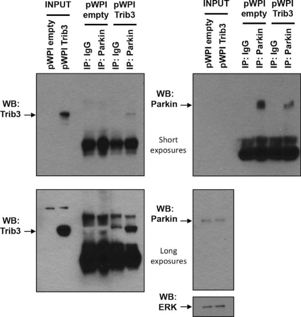
Figure 8.
Trib3 and Parkin physically interact in neuronal PC12 cells. Top, Short exposures. Bottom, Long exposures. Left panels, Western immunoblot images showing Trib3. Right panels, Parkin and ERK protein levels. Neuronal PC12 cells were transduced with an empty control vector (pWPI empty) or a Trib3-expressing vector (pWPI Trib3) for 48 h, and an immunoprecipitation (IP) experiment was performed with anti-Parkin antibody or control IgG.

