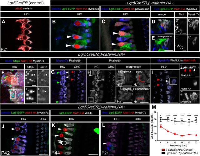
Figure 3.
Lgr5+ cells derived new HCs remain immature. A–L, Confocal images of immunostaining with multiple markers in cochleae from Lgr5CreER (Control) (A) and Lgr5CreER;β-catenin;HA+ (B–I) mice that were induced with tamoxifen at P0-P1 and analyzed at P21. A, IHCs express otoferlin and vGlut3 in control mice. New HCs (HA+) adjacent to the IHCs express Myosin7a (arrowheads in B and asterisk in D″), otoferlin (B, arrowheads), parvalbumin (C, arrowheads), but not the terminal IHC marker vGlut3 (C, arrowheads). In the new HCs, Tuj1 projections were present on the basolateral side of the ectopic HCs (D and arrowheads in D′). D–D″, Asterisk indicates the ectopic HC. In the endogenous IHCs, the synaptic markers Ctbp2 and GluR2 are both expressed in a matching pattern (arrowheads in F2′ and F2″, respectively). In the new HCs, some GluR2 puncta overlap with Ctbp2 puncta (arrowheads in F1′ and F1″, respectively), whereas others do not (open arrowhead in F1′ and F1″, respectively). G, The hair bundles form in the new HCs abutting the IHCs (green arrowheads in G), but their morphology differs from that of the endogenous IHCs (white arrowheads in G) and OHCs (red arrowheads in G). H, Phalloidin staining of the IHC region from G. Relative to the endogenous HCs (H1), the medially located new HCs display bent (H2), splayed (H3), or perpendicular morphology (H4). I, The ectopic HCs located in the OHC region (white arrowhead in I and I′) also display a peculiar hair cell bundle morphology, which does not resemble that of the traditional “V” shape found in normal endogenous OHCs (red arrowheads in I). I′–I″′, Cross-sectional view in which there is accumulation of actin at the cuticular plate in the endogenous (red arrowheads in I′) and ectopic HCs (white arrowhead in I′). J, K, New HCs are still present at P42-P44 (514.8 ± 18 per cochlea). Immunostaining of otoferlin (blue), Lgr5-EGFP (green), Atoh1-HA (red), Myosin7a (white in J), and vGlut3 (white in K) in the cochlea of a Lgr5CreER;β-catenin;HA+ mouse at P42-P44. L, New HCs generated from β-catenin and Atoh1 with BrdU pulse at P4 is still present at P21. M, ABR thresholds (± SEM) from P21 animals. Scale bars, 25 μm. Scale bars: D′, D″, E′, E″, H1–H4, I′–I″′, 10 μm; F, 5 μm; F1′, F2″, 2 μm. *p < 0.05 (two-way ANOVA with Bonferroni correction). ***p < 0.0001 (two-way ANOVA with Bonferroni correction). Refer to text for actual p values. N = 9 (control: β-catenin;HA+) and N = 6 (Lgr5CreER; β-catenin;HA+).

