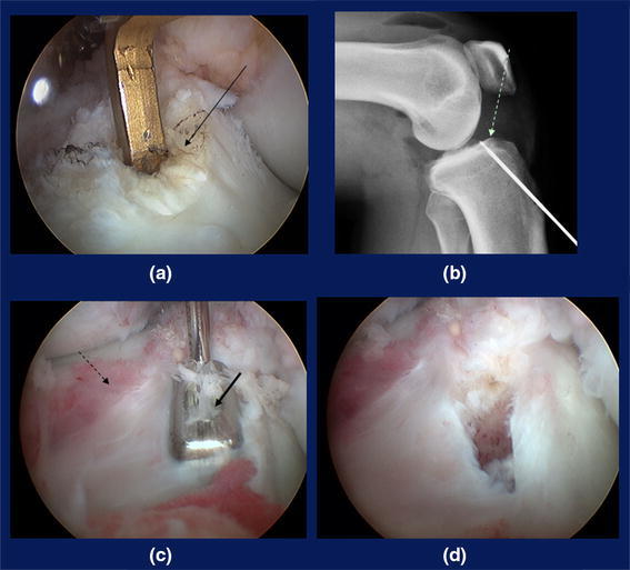
Fig. 9.

Tibial tunnel creation. a The tip aimer at the center, b Lateral radiograph showing the central pin and Parson’s Knob (dotted arrow). c The dilator (solid arrow) and the anterior horn of the lateral meniscus (dashed arrow). d Rectangular tunnel
