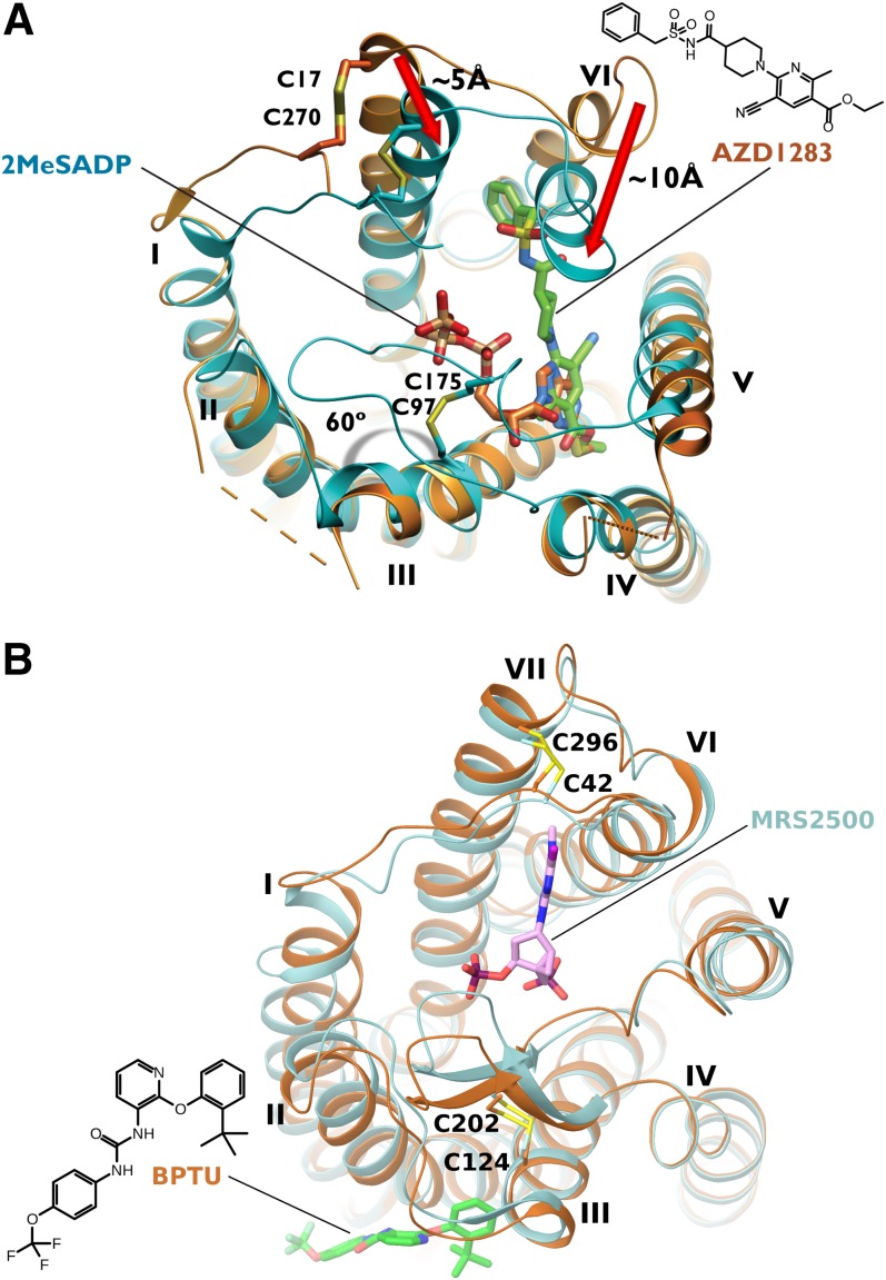
Fig. 4.
(A) Human P2Y12R X-ray structures in complex with AZD1283 (the non-nucleotide antagonist is shown in green carbon sticks, and the receptor is shown in orange ribbons) and 2MeSADP (the nucleotide full agonist is shown in orange carbon sticks, and the receptor is shown in cyan ribbons) (Zhang et al., 2014a,b). (B) Human P2Y1R X-ray structures in complex with the antagonists MRS2500 (the nucleotide antagonist is shown in pink carbon sticks, and the receptor is shown in cyan ribbons) and BPTU (the allosteric antagonist is shown in green carbon sticks, and the receptor is shown in orange ribbons) (Zhang et al., 2015).

