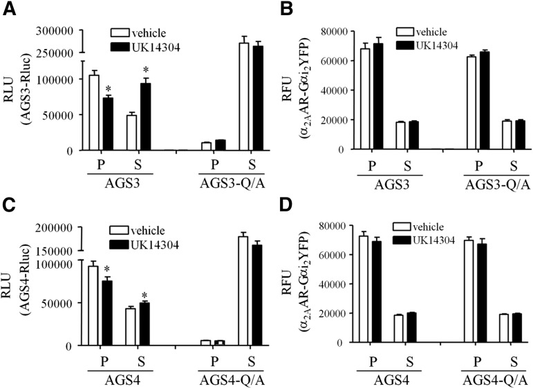
Fig. 3.
Agonist-induced changes in GPR protein distribution. AGS3Rluc (A and B) or AGS4Rluc (C and D) and α2AAR-Gαi2YFP were expressed in HEK293 cells as described in Materials and Methods. Cells were incubated with vehicle (Tyrode’s solution) or UK14304 (10 μM) for 5 minutes followed by hypotonic lysis, and AGS3Rluc (A) or AGS4Rluc (C) relative luminescence units (RLU) and α2AAR-Gαi2YFP relative fluorescence units (RFU) (B and D) were measured in supernatant (S) and pellet (P) fractions representing crude cytosol and membrane fractions, respectively. AGS3-Q/A and AGS4-Q/A refer to the mutation of a conserved glutamine residue in each of the GPR motifs to alanine, which renders them incapable of binding Gαi. *P < 0.05 compared with vehicle.

