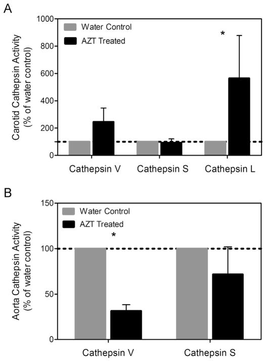
Fig. 7.
AZT alters cathepsin L activity. The level of activity of cathepsins S and L was assessed using zymography. (A) Densitometric analysis of the carotids revealed an increase in cathepsin L, while analysis of the aortas showed decreases in cathepsin L activity. (B) (n = 4, p < 0.05, and data is mean ± SEM).

