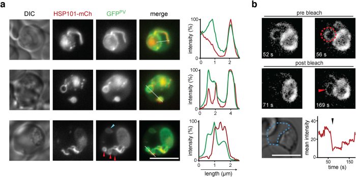
Figure 3. HSP101 delineates a tubular subcompartment of the parasitophorous vacuole.
(a) Live co-localization of HSP101-mCherry (centre left) with a marker protein of the parasitophorous vacuole (GFPPV, centre). The line in the merge (centre right) indicates profiling of the fluorescent signal (right). Shown are three representative trophozoites demonstrating vacuolar tubules, loops, and vesicles. The blue arrowhead denotes a detached, vacuole-derived, and HSP101-mCherry negative lumen. The red arrowheads denote budding structures at the site of a vacuolar tubule. Note that HSP101-mCherry is excluded from these compartments. (b) FRAP analysis reveals free diffusion from the parasitophorous vacuole to the tubular extensions. Erythrocytes infected with mCherryPV parasites were analysed by confocal microscopy before (pre bleach) and after (post bleach) photo bleaching (red area, bleach location). Shown is a representative trophozoite and the respective temporal fluorescence analysis in the erythrocyte cytoplasm (blue dotted line); black arrowhead indicates time of the bleaching pulse. Scale bar, 5 μm.

