
Figure 4. APT analysis of the most heavily doped sample Ce0.2Co4Sb12.

(a) 3D reconstruction of microtip containing a grain boundary. Ce atoms are displayed in red; Sb and Co atoms omitted for clarity. (b) Concentration profile across the grain boundary and in the grain. The black dashed lines show values measured by EPMA, and the error bars represent the s.e., 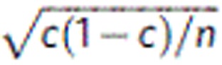 , where c is the concentration and n is the number of atoms detected in each data point.