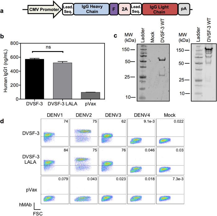
Figure 1. In vitro expression of human anti-DENV neutralizing mAbs by DMAb.
(a) Schematic illustration of DNA plasmid used for DMAb; antibody heavy and light chain sequences are separated by a combination of furin and 2A cleavage sites. (b) ELISA quantification analysis of human IgG in supernatants of pDVSF-3 WT- or LALA-transfected 293T cells. The data displayed are the mean of triplicate values +/− standard error of the mean (SEM) and are representative of three independent experiments. (c) Western blot analysis of pDVSF-3 WT-transfected 293T supernatants containing DVSF-3 WT. Antibodies were purified by Protein A spin columns and separated by SDS-PAGE under reducing (left) and non-reducing (right) conditions.(d) Vero cells were either uninfected (Mock) or infected by DENV1, 2, 3, or 4, then fixed, permeabilized, and stained with supernatants of pDVSF-3 WT- or LALA-transfected 293T cells. The data displayed are representative of two independent experiments.

