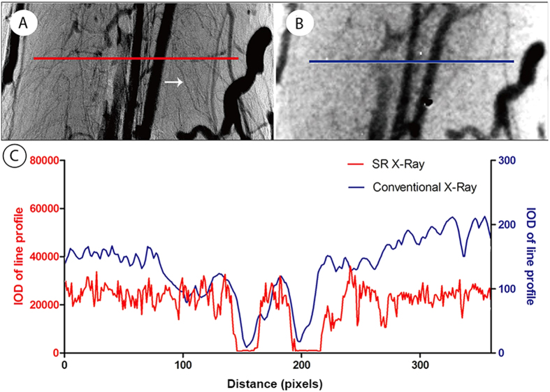
Figure 2. Comparison of initial projected images between synchrotron radiation (SR) and conventional X-ray imaging.
(A) Projected image by synchrotron radiation imaging. (B) Projected image by ordinary X-ray imaging. (C) Representation of the intensity distribution of the line profiles located in the correspondence images in B and C indicating that the SR (red line) has a greater capacity to distinguish the microvessels.

