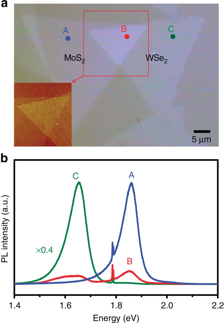
Figure 1. Optical microscopy images and photoluminescence spectroscopy taken on the staked WSe2/MoS2 heterostructure.
(a) Optical micrograph and atomic force microscopy images for the WSe2/MoS2 heterostructural stacked flakes on a sapphire substrate. (b) Photoluminescence spectra for the selected sites including MoS2 only (A), WSe2 only (C) and WSe2/MoS2 (B) stacked areas.

