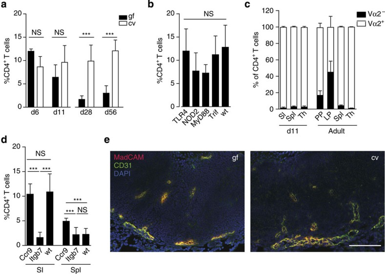
Figure 3. Postnatal homing of CD4 T cells to the neonatal intestine.
(a) Comparative analysis of the percentages of CD4+TCRβ+ T lymphocytes among total CD45+ immune cells in intestinal tissue of conventional (cv) and germ-free (gf) mice at the indicated age (n=8–11 from two experiments, mean±s.d.; one-way ANOVA, Bonferroni's post test, ***P<0.001; NS, not significant). (b) Comparative analysis of the percentage of CD4+TCRβ+ T lymphocytes among total CD45+ immune cells in wild type (wt), Tlr4−/−, TrifLps2/Lps2, MyD88−/− and Nod2−/− mice at 6 days after birth (n=4–11, mean±s.d.; one-way ANOVA, Bonferroni's post test; NS, not significant). (c) Comparative analysis of the frequency of Vα2+ and Vα2− cells among TCRβ+CD4+ lymphocytes in the total small intestine (SI), spleen (Spl) and thymus (Th) of 12-day-old and PPs, LP, spleen (Spl) and thymus (Th) of adult OTII transgene mice (n=7 and 3, respectively; representative of two independent experiments, mean±s.d.). (d) Comparative analysis of the percentage of CD4+TCRβ+ T lymphocytes among total CD45+ immune cells in the small intestine (SI) and Spleen (Spl) of wild type (wt), Ccr9−/− and Itgb7−/− mice at 6 days after birth (n=7–11 from two experiments, mean±s.d.; one-way ANOVA, Bonferroni's post test, ***P<0.001; NS, not significant). Note that the same data set from the neonatal wt/cv group is shown in a–c because the figures represent pooled data. (e) Immunofluorescence staining of MadCAM-1 (red) in combination with CD31 (green) on PP tissue sections of gf and cv 11-day-old neonates dissected using the binocular. Magnification, 1 × 200; counterstaining with DAPI (blue; scale bar, 100 μm; n=1, representative of two experiments).

