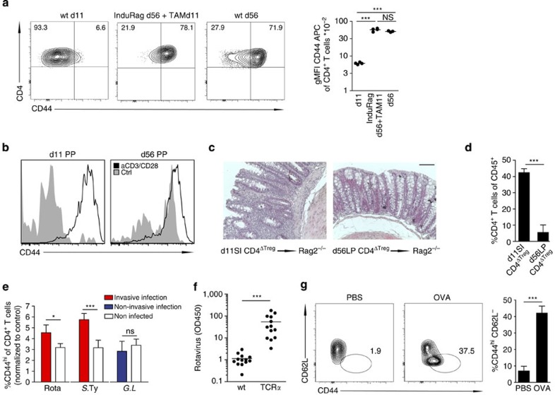
Figure 4. Maturation of neonatal CD4 T lymphocytes following mucosal challenge.
(a) Lymphocyte maturation status in the adult lymphopenic host after the induction of Rag in InduRag mice. FACS analysis (left panel) and gMFI of CD44 (right panel) on CD4+TCRβ+ lymphocytes obtained from the SI of 11- and 56-day-old wt (wt d11 and wt d56) and adult InduRag mice 11 days after Tamoxifen administration (InduRag d56+TAM11). (n=3–4, representative of two experiments, mean; one-way ANOVA, Bonferroni's post test, ***P<0.001; NS, not significant). (b) Ex vivo activation of CD4+TCRβ+ lymphocytes from PP of 11- and 56-day-old mice after 3 days of culture in presence of anti-CD3/CD28 beads (n=4, one experiment). (c) H&E staining and (d) percentage of infiltrating CD4+TCRβ+ lymphocytes of CD45+ in the colonic tissue of adult Rag2 recipients 5–6 weeks after transfer of 1–5 × 10⁴ SI CD4+ T cells from 11-day-old or adult Foxp3-GFP donor mice after TReg depletion (T-cell transfer colitis model). Magnification, × 40 (bar graph 100 μm). (n=3–4, representative of two experiments, mean±s.d.; unpaired Student's t-test, ***P<0.001). (e) Percentage of CD44hi cells among CD4+TCRβ+ lymphocytes (using the 3% of CD4 T cells in the non-infected control mice with the highest CD44 expression as a reference gate) in the neonate SI after infection with rotavirus (infected at d4 post parturition, pp and analysed at 8 d.p.i.), S. Typhimurium (infected at d4 pp and analysed at 4 d.p.i.) and Giardia lamblia (infected d4 pp and analysed at 8 d.p.i.). (One litter (n=6–11) from the infected group and four age-matched non-infected controls were analysed per experiment; representative of two experiments, mean±s.d.; unpaired Student's t-test, *P<0.05, ***P<0.001; NS, not significant). (f) Quantification of rotavirus antigen by ELISA in colon homogenate of wt and TCRα−/− neonates at 8 d.p.i. infected at 4d pp (n=12–13 from two experiments, mean; Mann–Whitney U-test, ***P<0.001). (g) FACS plots (left panel) and percentages (right panel) of CD44hiCD62L− of CD4+TCRβ+ lymphocytes in PPs. 11-day-old DO11.10 neonates were gavaged daily with 10 mg OVA or PBS starting at d3 pp (n=5, representative of two experiments, mean±s.d.; unpaired Student's t-test, ***P<0.001).

