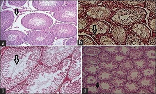
Figure 8.

Effects of Mn2O3 nanoparticles over the damage of testis. (a) Arrows show elevation in cellular disruption of seminiferous tubules, interstitial edema, and decline in cell regulation were observed by H and E (×10). (b) Arrows show chaos in the germinal cells level in seminiferous tubules, increased in the gap between seminiferous tubular, and vacuoles seen in epithelium by H and E (×10). (c) Arrows indicate elevation in diameter of seminiferous tubules and decline in epithelium diameter were observed, H and E (×10). (d) Arrows indicate images of seminiferous tubules in the control-group demonstrated uniformity of the seminiferous tubules were seen, H and E (×10)
