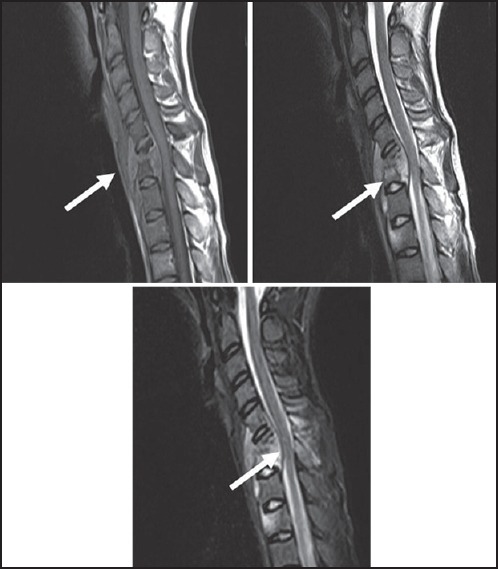
Figure 1.

Tubercular Spondylodiscitis — Sagittal T1, T2 and STIR images show anterior wedging of C6 and C7 vertebral bodies. Disc at C6-7 level is inappreciable on T1 and T2W images. Subligamentous spread (from C5-D2) and anterior epidural soft tissue(C5-7) is seen
