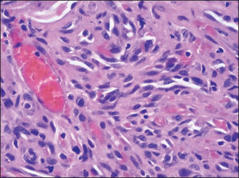
Figure 2.

Photomicrograph of the histopathologic section reveals fibrous connective tissue stroma with numerous irregular slit like spaces containing extravasated red blood cells and surrounded by ill-defined fascicles of spindle shaped cells (H-E)

Photomicrograph of the histopathologic section reveals fibrous connective tissue stroma with numerous irregular slit like spaces containing extravasated red blood cells and surrounded by ill-defined fascicles of spindle shaped cells (H-E)