
Figure 2.
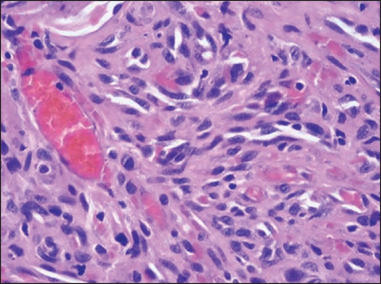
Photomicrograph of the histopathologic section reveals fibrous connective tissue stroma with numerous irregular slit like spaces containing extravasated red blood cells and surrounded by ill-defined fascicles of spindle shaped cells (H-E)
Official websites use .gov
A
.gov website belongs to an official
government organization in the United States.
Secure .gov websites use HTTPS
A lock (
) or https:// means you've safely
connected to the .gov website. Share sensitive
information only on official, secure websites.
Photomicrograph of the histopathologic section reveals fibrous connective tissue stroma with numerous irregular slit like spaces containing extravasated red blood cells and surrounded by ill-defined fascicles of spindle shaped cells (H-E)