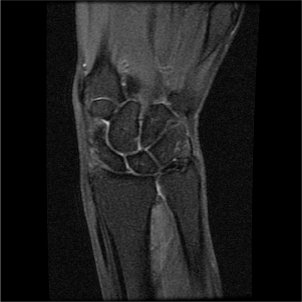
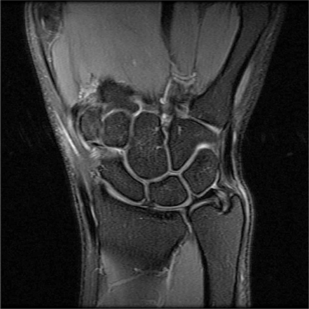
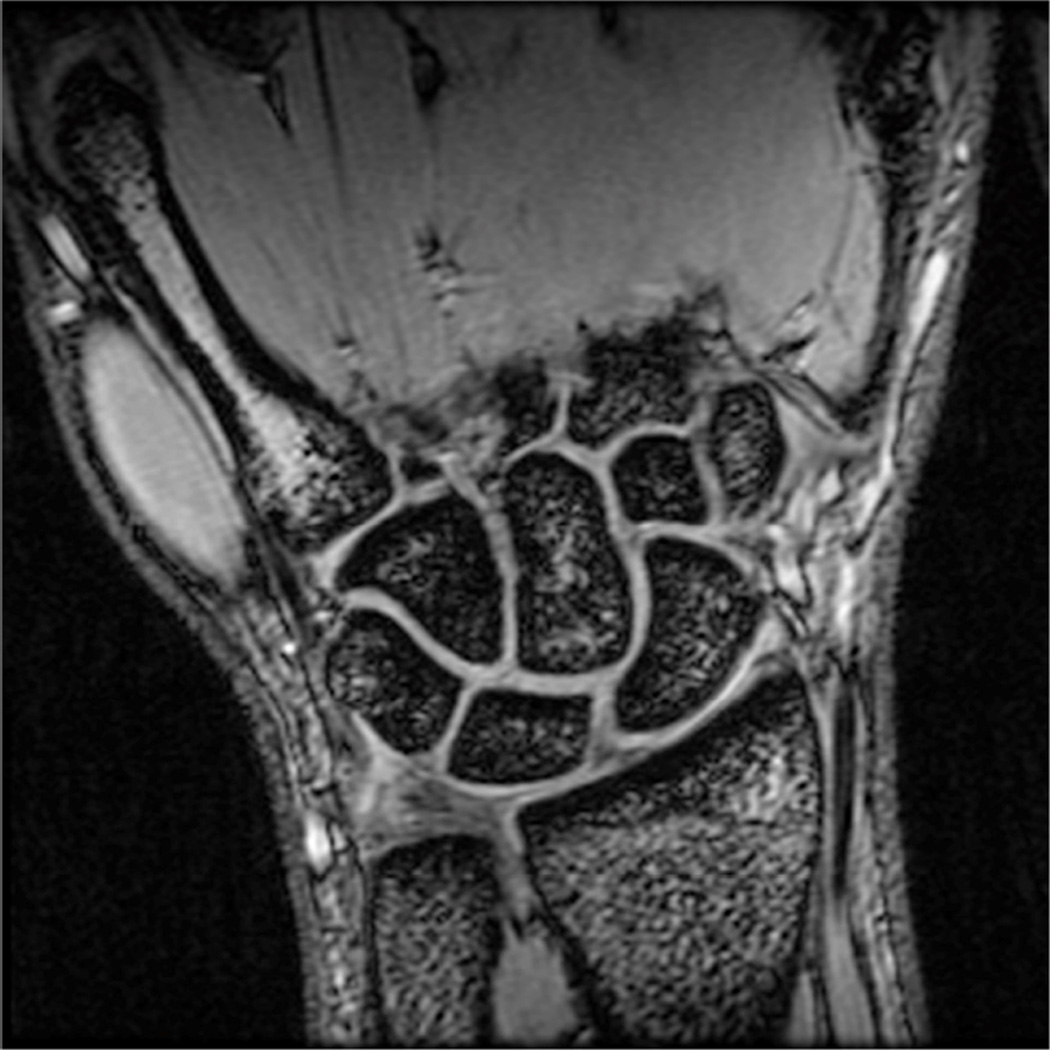
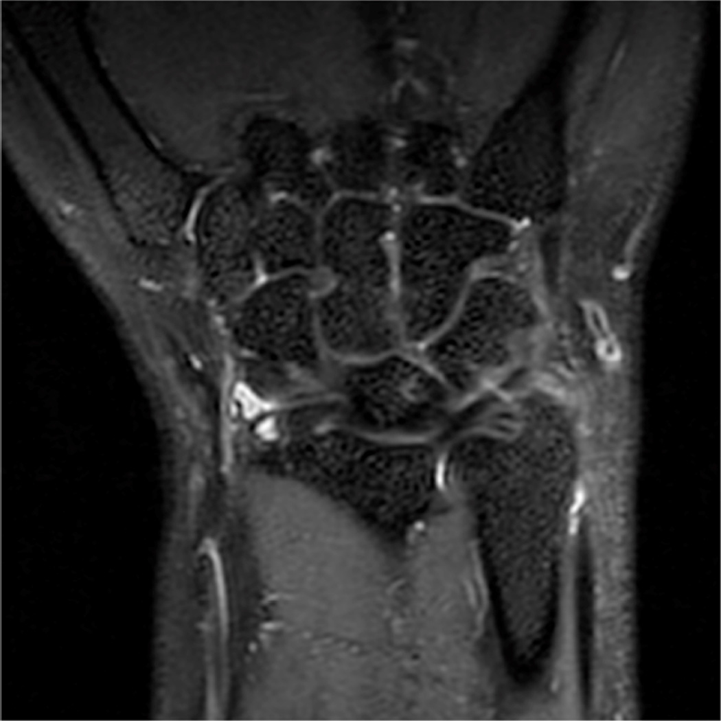
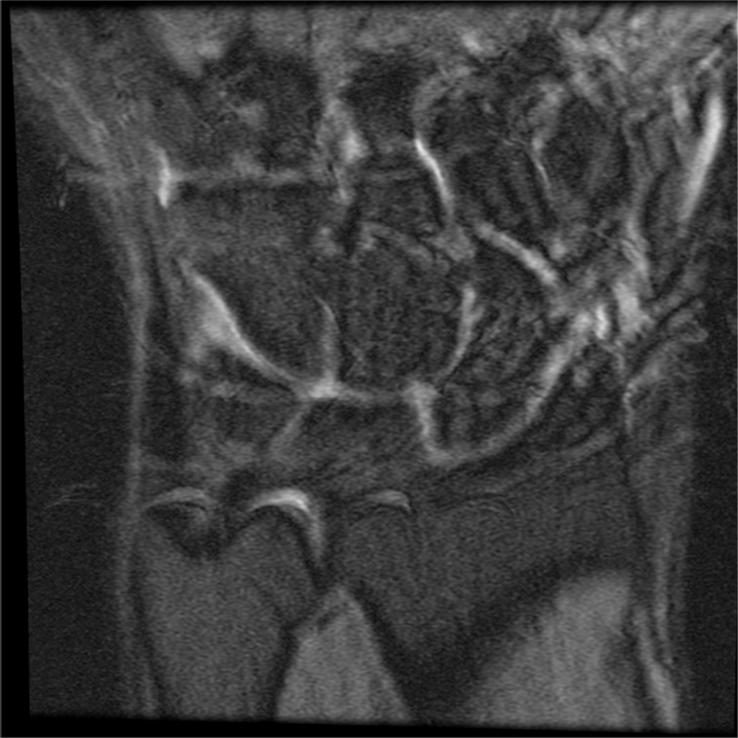
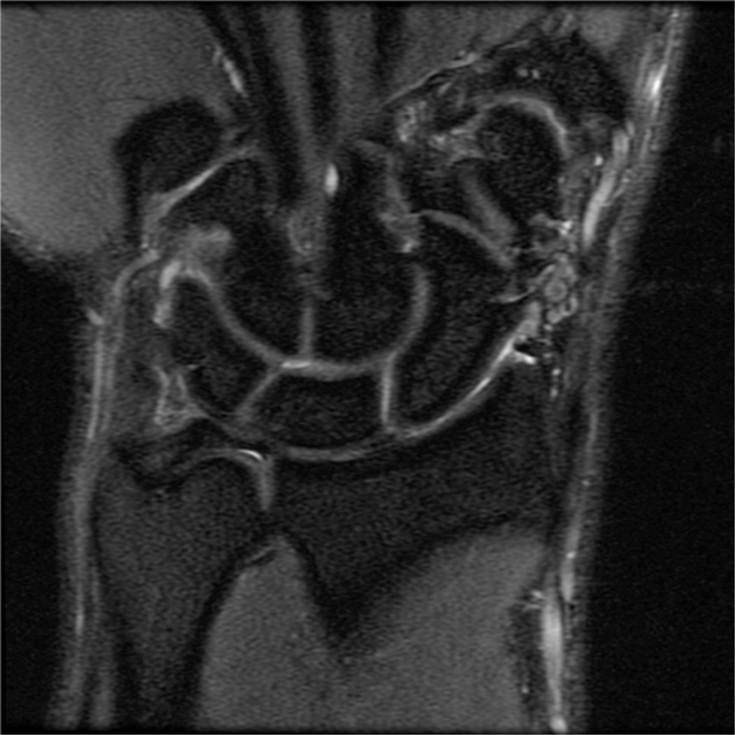
Figure 11.
Representative images of not recommended and recommended MR imaging protocols. Low-resolution not recommended (A) coronal image of the wrist using a 16 cm FOV and 224×128 matrix, 3 mm slice thickness. High-resolution recommended protocol (B) using a 10 cm FOV, 384×192 matrix and 2 mm slice thickness. Coronal 3D gradient echo acquisition (C) is inferior for bone marrow lesions and ligament tears and not recommended compared with newer 3D spin-echo based methods such as 3D FSE Cube (D). Uncomfortable positioning of the joint can lead to subject discomfort and motion (E), which can be corrected with repositioning the joint in a more comfortable location (F).

