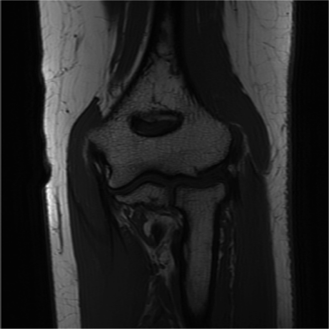
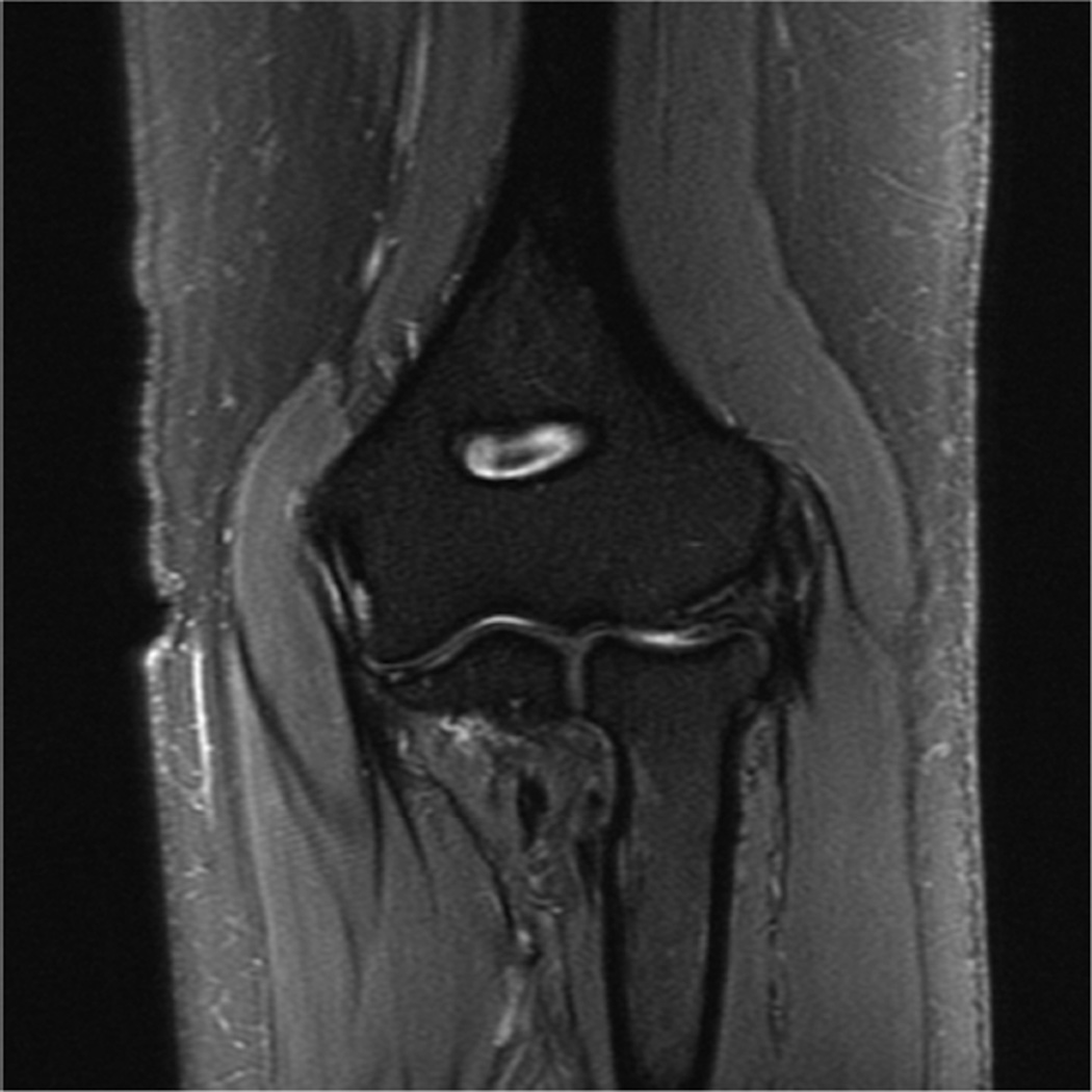
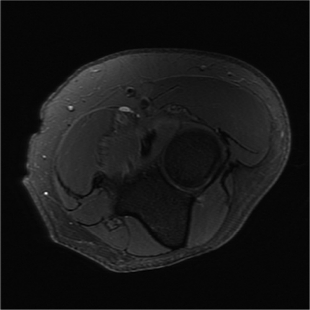
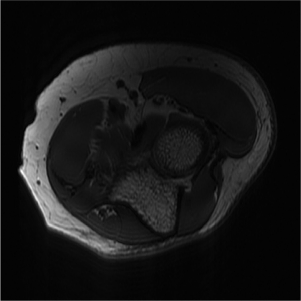
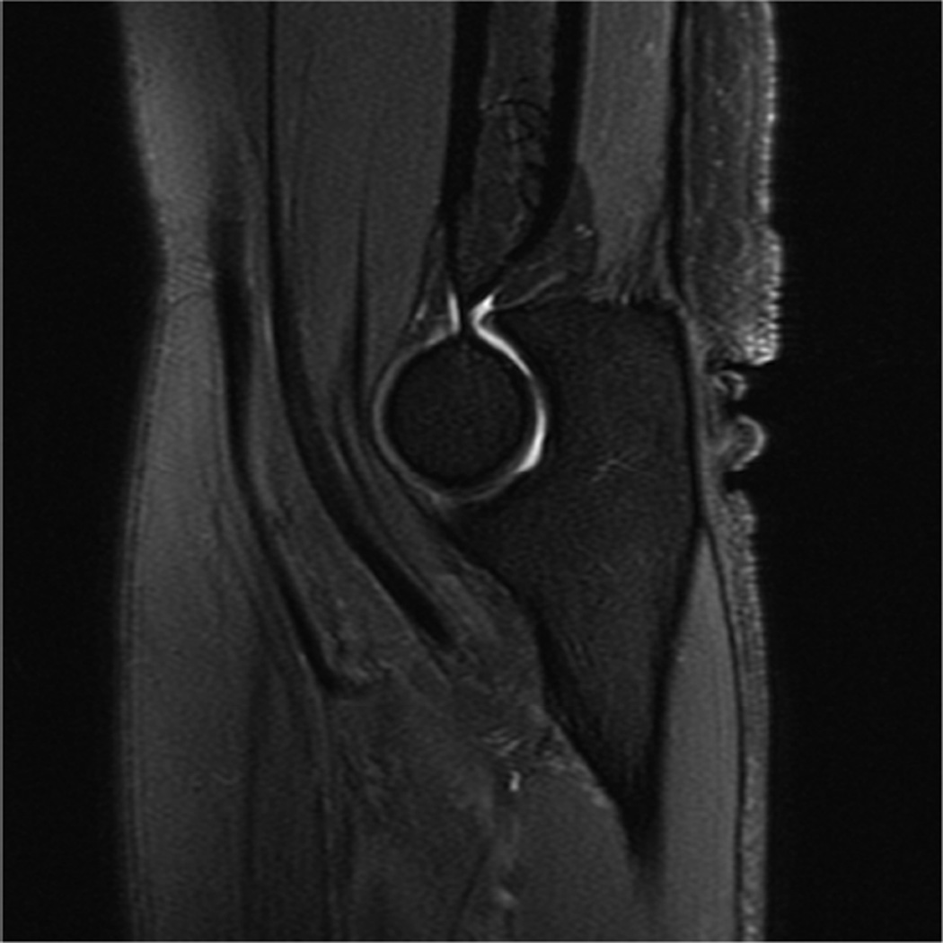
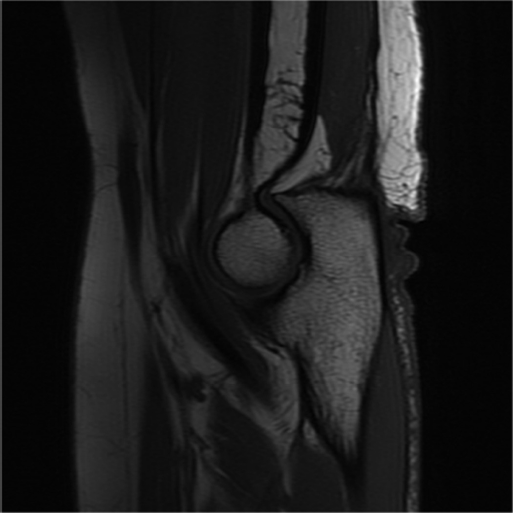
Figure 6.
Images from the 3T elbow protocol shown in Table 3, acquired in a patient with the 16-channel phased array flex coil. A) Coronal T1-weighted B) Coronal proton-density-weighted (PD) with fat saturation (FS) C) Axial PD FS D) Axial T1-weighted E) Sagittal PD FS F) Sagittal T1- weighted.

