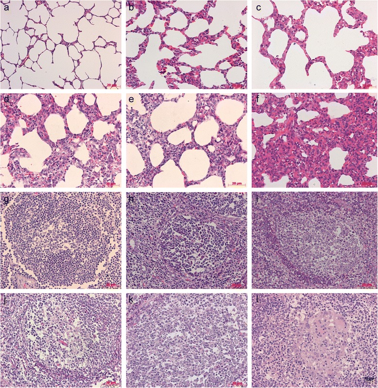
Fig. 5.

Histopathological lesions in experimental conventional pigs. a No markable microscopic lesions in the lung of mock group pigs. b Slight lymphoplasmacytic and histiocytic bronchointerstitial pneumonia in the lung of a pig vaccinated with 2×104.0 TCID50 dose attenuated PCV1-2b. c Mild lymphoplasmacytic and histiocytic bronchointerstitial pneumonia in the lung of a pig vaccinated with inactivated PCV1-2b. d Moderate lymphoplasmacytic and histiocytic bronchointerstitial pneumonia in the lung of a pig vaccinated with 2×103.5 TCID50 dose attenuated PCV1-2b. e Conspicuous lymphoplasmacytic and histiocytic bronchointerstitial pneumonia in the lung of a pig vaccinated with commercial inactivated PCV2b vaccine X. f Severe lymphoplasmacytic and histiocytic bronchointerstitial pneumonia in the lung of a nonvaccinated challenged pig. g No remarkable microscopic lesions in the lymph nodes of a pig in the mock group. h Slight lymphoid depletion (LD) in lymph node follicles of a pig vaccinated with 2×104.0 TCID50 dose attenuated PCV1-2b. i Mild LD in lymph nodes follicles of a pig vaccinated with inactivated PCV1-2b. j Moderate LD in lymph nodes follicles of a pig vaccinated with 2×103.5 TCID50 dose attenuated PCV1-2b. k Conspicuous LD in lymph nodes follicles of a pig vaccinated with commercial inactivated PCV2b vaccine X. l Moderate histiocytic replacement (HR) in lymph node follicles of a nonvaccinated challenged pig. Bar = 20 μm (400×)
